
Cellular ageing is often spoken about as though it were a single clock ticking down…
Six Organs, One Discovery: Testes, Kidney, Brain, Thymus, Retina and Heart Stem Cell Secretomes Show Anti-Senescent Potential
Cellular ageing is often spoken about as though it were a single clock ticking down. At the microscopic level, it is more like a chain reaction: stress accumulates, important molecules are damaged, DNA alarms are triggered, and cells can enter a state in which they remain alive but no longer function as well as they should. In the brain, that matters because long-lived nerve cells must cope with stress for decades.
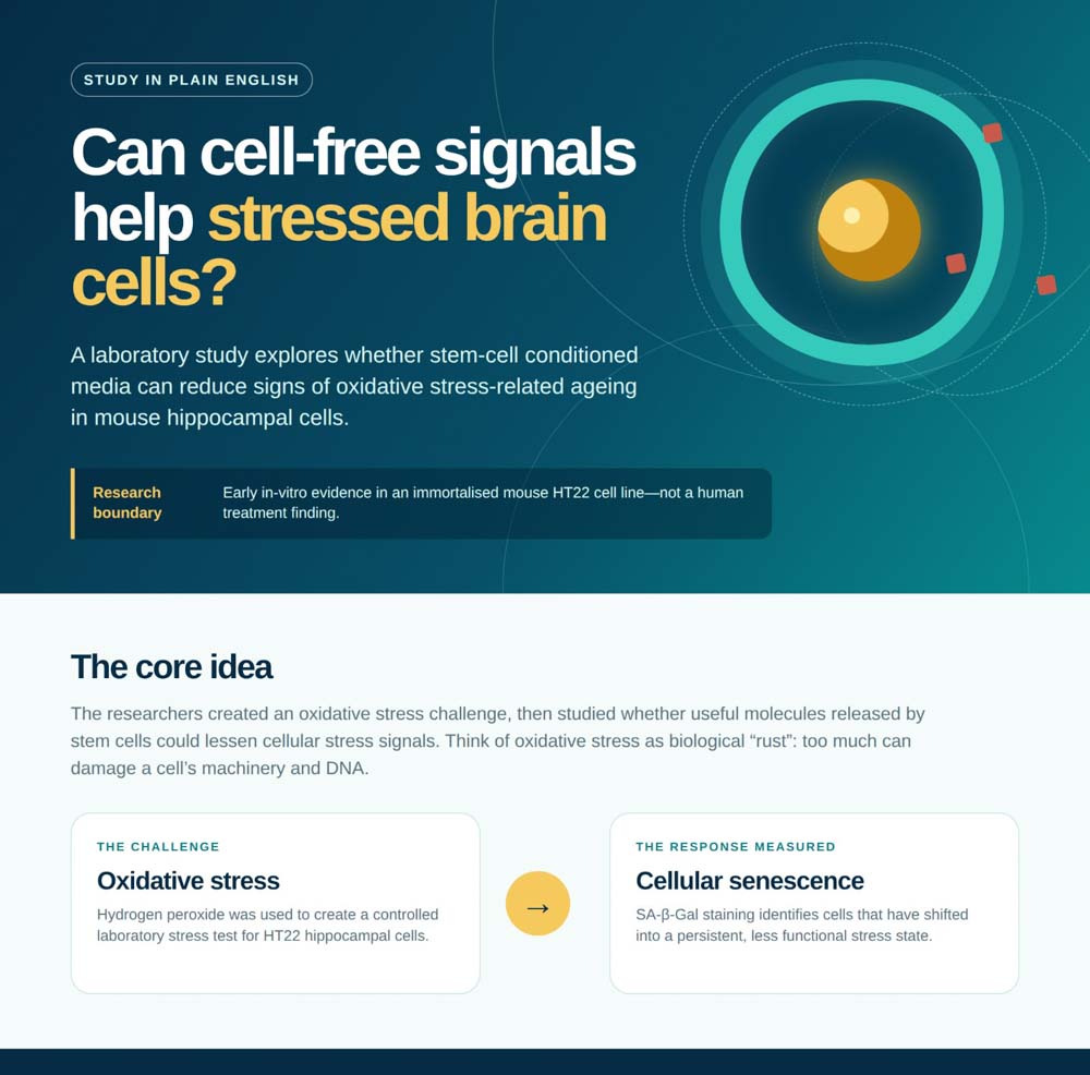
The study “Organ-Specific Stem-Cell Conditioned Media Attenuate Oxidative Stress-Induced Senescence and DNA Damage in HT22 Hippocampal Neuronal Cells” examines an intriguing possibility: could cell-free signals released by targeted organ-specific precursor stem cells lessen stress-induced senescence in neuronal cells without transplanting living stem cells themselves?
The short answer is yes, in this controlled laboratory model. All six organ-specific precursor stem-cell conditioned-media preparations reduced a marker of cellular senescence, and all showed a visible reduction in a DNA-damage marker. This is an early, targeted organ-specific anti-senescence signal in an immortalised mouse hippocampal cell line—not evidence of a treatment for patients or a clinical recommendation.
Study at a Glance
The infographic below brings together the study’s laboratory pathway, the six reported organ-specific results, Prof. Mike Chan’s targeted organ-specific exosome/secretome research strategy and the key boundaries on what these early findings can—and cannot—show.
Why Cellular Aging Matters
Think of oxidative stress as biological “rust.” Just as metal can corrode when exposed to the elements, cells can be harmed by an excess of reactive oxygen species. In this experiment, the researchers used hydrogen peroxide (H₂O₂) to create that stress in HT22 cells, a commonly used mouse-derived hippocampal neuronal cell model.
The stress signal was strong. The average number of SA-β-Gal-positive cells—a standard laboratory indicator of senescence—rose from 2.3 in untreated controls to 535.7 after oxidative stress. That large separation matters because it shows the test system was capable of detecting whether an intervention made a difference.
Two complementary checks were used:
- SA-β-Gal staining identifies cells that have entered a senescent, less functional state. It is similar to seeing a warning light that tells us a cell has shifted into a persistent stress mode.
- γH2A.X foci are small fluorescent signals that appear around DNA double-strand breaks. They are like markers placed around a damaged section of a document to show where repair attention is needed.
Using both measures is one of the study’s strengths. A reduction in one marker alone can be misleading; seeing a quantitative fall in senescence alongside a qualitative reduction in DNA-damage signals creates a more coherent biological picture.
“The value of this work is not a promise to patients; it is a practical clue that stressed nerve cells may respond to cell-free protective signals.”
What the Study Actually Found
The researchers collected conditioned media—the nutrient fluid carrying the cell-free cargo released by targeted organ-specific precursor stem cells—from six tissue sources: retina, kidney, brain, testes, thymus and heart. Each sample was therefore an organ-specific preparation. This fluid is often called the secretome. Rather than delivering living cells, the approach investigates the proteins, growth factors, antioxidant enzymes, extracellular vesicles—including exosomes—and other messages that cells leave behind.
A useful everyday comparison is a repair crew and its toolkit. A live-cell transplant brings the whole crew into a new building. A targeted organ-specific cell-free approach aims to deliver useful tools and instructions from a defined precursor-cell source—without bringing in the cells themselves. The experiment tested the complete conditioned media, so it did not establish that exosomes alone, or any one molecule, caused the observed effect. Identifying the active exosome/secretome cargo is therefore an important next research step.
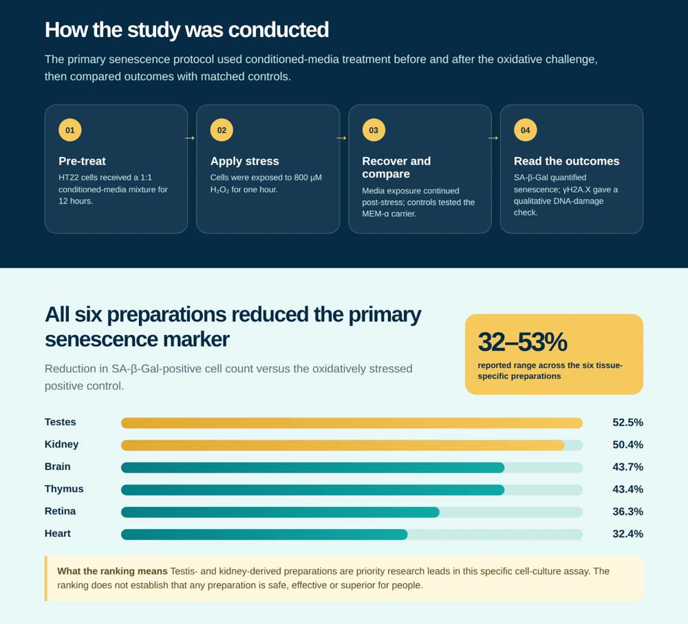
Every targeted organ-specific conditioned-media preparation reduced SA-β-Gal-positive cells relative to the stressed control, with statistically significant results in this experiment. This supports a comparative anti-senescence signal across the six preparations in the HT22 hippocampal cell assay; it does not demonstrate anti-senescence of entire organs. The reductions were:
| Tissue source | Reduction in senescence marker | Plain-language interpretation |
|---|---|---|
| Testes | 52.5% | Strongest result in this laboratory comparison |
| Kidney | 50.4% | Close second and a priority research lead |
| Brain | 43.7% | Intermediate protective signal |
| Thymus | 43.4% | Intermediate protective signal |
| Retina | 36.3% | Meaningful but smaller reduction |
| Heart | 32.4% | Smallest reduction, still significant in this model |
The testis- and kidney-derived preparations led the ranking. This indicates the strongest signals from those organ-specific precursor-cell preparations in this particular model and assay. It does not mean these tissues are proven “best” for people, or that a product from them should be used clinically; it means they are sensible candidates for the next round of mechanism and validation work.
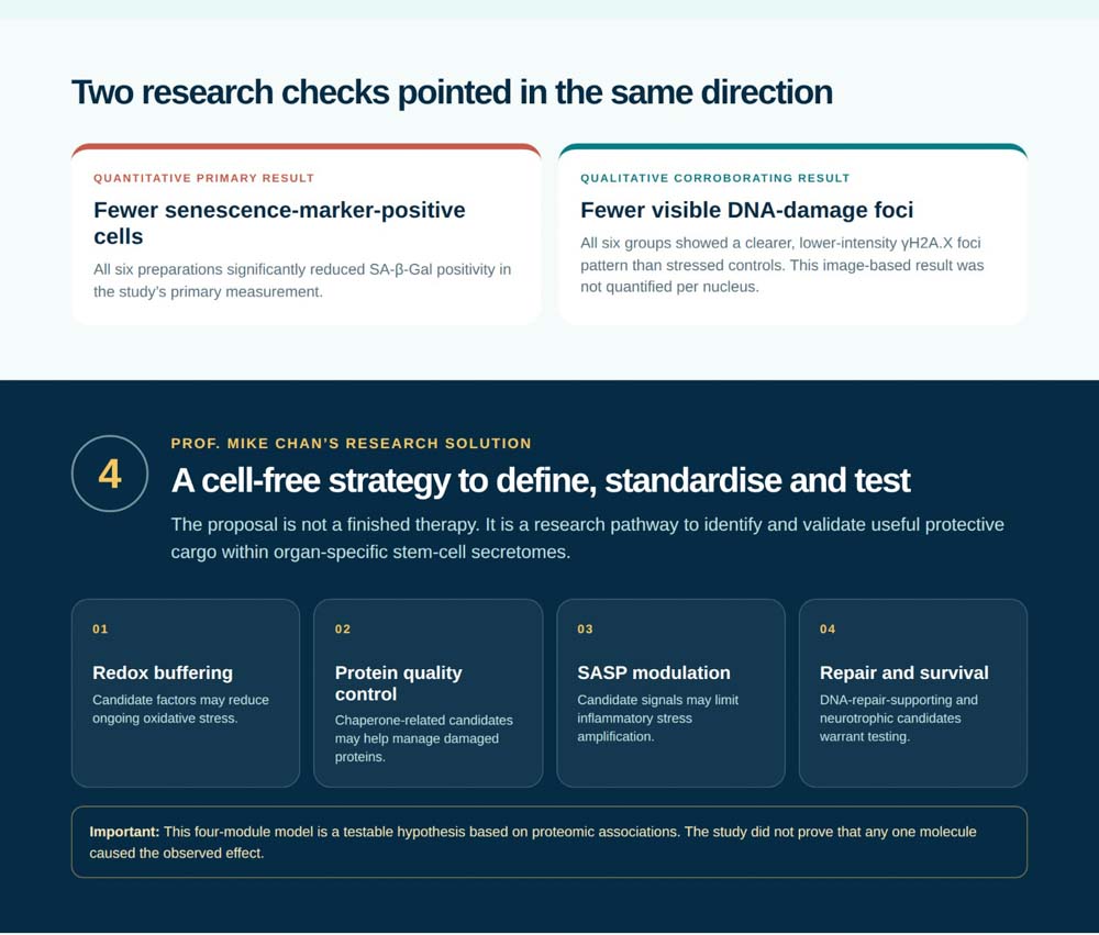
The DNA-damage images told a compatible story. All six preparations showed fewer and dimmer γH2A.X foci than the stressed control. Importantly, this measurement was qualitative—the paper did not report a numerical count of foci per nucleus—so the finding supports, but does not quantify, a reduction in DNA-damage burden.
Another detail strengthens the interpretation. The culture carrier medium, MEM-α, had no significant effect on stressed or unstressed controls. This makes it more plausible that the benefit came from molecules and vesicles released by the organ-specific precursor stem-cell cultures rather than the carrier fluid itself. It does not, however, distinguish exosomes from the other components of the whole conditioned media.
“When different readouts point in the same direction, we gain a stronger research signal—but we still need to test the signal in more realistic biological settings.”
The proteomic findings also help explain why the response may not be due to one miracle molecule. Across the six secretomes, the study identified 754 proteins, with 359 detected in all six. The authors propose a combined, testable four-part protective model:
- Redox buffering may help neutralise residual oxidants, much as a spill kit helps contain a corrosive leak.
- Protein quality control may help stressed cells manage damaged or misfolded proteins.
- Inflammation and SASP modulation may help prevent stressed cells from amplifying inflammatory signals to their neighbours.
- DNA-repair support and neurotrophic signalling may help cells recover from damage and maintain survival pathways.
This four-part explanation remains a hypothesis, not a confirmed mechanism. The study identifies plausible candidate molecules—including peroxiredoxins, glutathione peroxidase 3, chaperones, clusterin, IGF-related proteins and extracellular-vesicle markers—but did not yet remove or block them one at a time to prove which ones drive the effect.
Prof. Mike Chan’s Targeted Organ-Specific Research Roadmap
Based on the study, Prof. Mike Chan’s solution is a targeted organ-specific exosome/secretome research strategy derived from targeted organ-specific precursor stem cells, rather than a direct stem-cell transplantation strategy. In this publication, the assay used whole conditioned media—the complete cell-free cargo—so exosomes are a central candidate component but are not yet proven to be the sole mediator. The near-term objective is not to offer a finished intervention; it is to turn an encouraging laboratory signal into a defined, reproducible and safe candidate for further study.
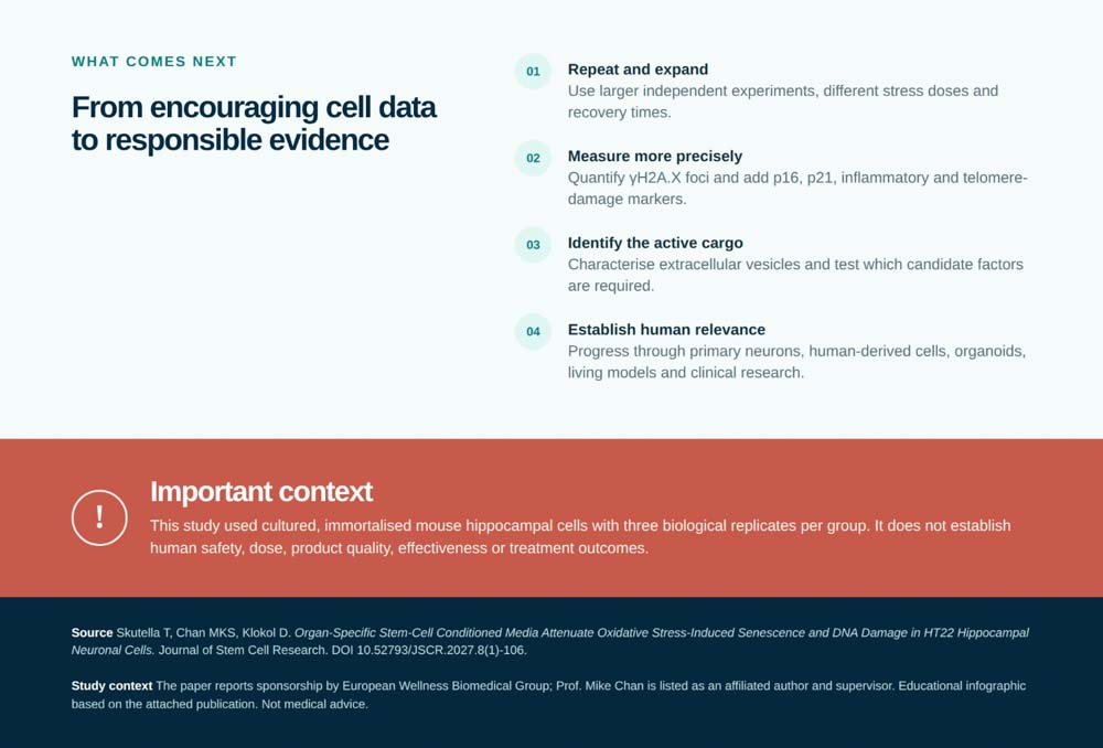
The paper points to several practical recommendations:
- Prioritise testis- and kidney-derived secretomes for deeper study. They achieved the largest reductions in the quantitative senescence marker, so they are logical starting points for identifying active components.
- Standardise the targeted organ-specific exosome/secretome cargo. A preparation is a mixture, not a single drug. Future batches should be profiled for protective protein groups, exosome and extracellular-vesicle content, particle number, size distribution and quality markers so that one preparation can be fairly compared with another.
- Test the proposed four-module model. The redox, chaperone, anti-inflammatory and DNA-repair/neurotrophic modules should be challenged experimentally. For example, selectively removing clusterin, HMGB1 or selected antioxidant activity could reveal whether a component is necessary, helpful or incidental.
- Strengthen the evidence with larger and more precise experiments. The current study used three biological replicates per group. Replication with larger samples, multiple stress doses and different recovery times would provide a clearer picture of consistency and dose range.
- Measure more than one type of cellular ageing. Future work should count γH2A.X foci per nucleus and add markers such as p16, p21, inflammatory SASP cytokines, and telomere-associated DNA damage. That is the difference between seeing a general warning sign and checking several gauges on the same dashboard.
- Move carefully toward human relevance. Before any patient-facing claim, results should be checked in primary neurons, human-derived neuronal cells, brain organoids and living models of ageing or neurodegeneration, alongside biodistribution, safety, purity and dosing studies.
This roadmap matters because an attractive biological story is not enough. A useful targeted organ-specific cell-free product must have a defined precursor-cell source, be consistently made, be carefully characterised—including its exosome and extracellular-vesicle content—be tested in human-relevant systems, and be proven safe and effective in appropriately designed clinical trials.
“The most responsible next step is not to overstate an early result, but to identify the active cargo, standardise it and test it where it matters most.”
Bottom line. This study provides a clear preclinical anti-senescence signal for a targeted organ-specific cell-free strategy: complete conditioned media from targeted organ-specific precursor stem cells lessened laboratory markers of oxidative stress-induced senescence in mouse hippocampal neuronal cells. Its most important contribution is the convergence of two indicators—one quantitative marker of senescence and one qualitative marker of DNA damage—together with a tissue-specific ranking that gives researchers a rational place to focus next. The study did not show a whole-organ effect in people, and it did not isolate exosomes to show that they are the sole cause of the response. The promise is real as a research direction; the responsible message is that it requires substantial validation before it can be translated into human care.
This commentary is educational and based solely on the attached publication. It is not medical advice and does not endorse, prescribe or recommend any stem-cell or secretome product.
Follow European Wellness:



Follow European Wellness Academy:
Follow Prof. Dato’ Sri Dr. Mike Chan:

Related Posts